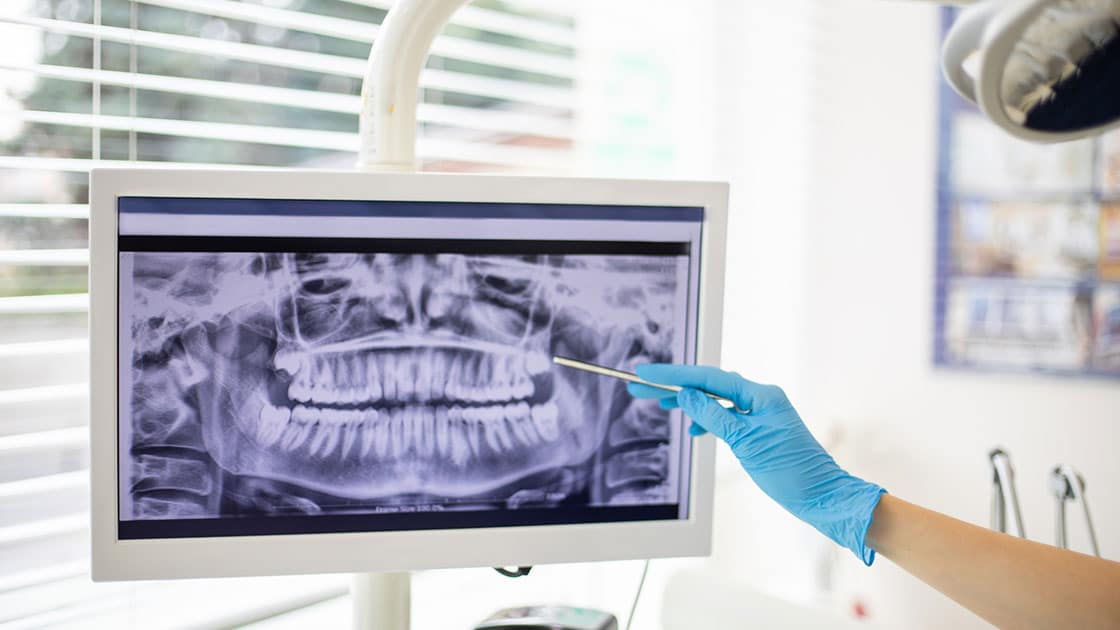
Cone Beam Scanner 3D Imaging
3D imaging provides better quality and more detailed images than traditional x-rays. This new technology is also less invasive and emits less radiation than traditional x-ray machines.
The landscape of oral health care has been transformed by the advancements in dental technology, presenting cutting-edge solutions that bolster patient treatment, elevate diagnostic precision, and streamline therapeutic procedures. Through breakthroughs in imaging technology that offer unparalleled insights into the mouth to the creation of robust and visually appealing materials for dental repairs, dental technology effectively connects the dots between professional clinical skill and the requirements of patients.
3D imaging provides better quality and more detailed images than traditional x-rays. This new technology is also less invasive and emits less radiation than traditional x-ray machines.
The GentleWave® Procedure takes a different approach to root canal therapy, using a minimally invasive protocol designed to help preserve optimal tooth structure and promote the long-term sustainability of the tooth.
In addition to being much more comfortable for the patient, using the scanner allows us to be more efficient and accurate, which can result in a reduced number of visits to the office, saving the patient time.
Digital x-rays reduce the amount of radiation needed as compared to film x-rays. The improved diagnostic capability of digital x-rays and the ability to view the x-rays on a computer screen allow the patient to better understand and follow treatment. Digital x-rays are instant, there is no longer a need to develop the film. Digital x-rays save time and increase patient care. It’s also a very green technology. By eliminating film, developer and chemical waste it is better for you and the environment!
The use of specialized operating microscopes means that the doctor is able to get a detailed look at the work they are doing during all phases of your endodontic treatment. The additional magnification and illumination allow them to work with great precision and see small details such as calcified canals and fractures. The Endodontist is able to more accurately diagnose and treat the patient using a dental surgical microscope to improve the potential outcome of the treatment from “good” to “excellent”. Further, some microscopes may be equipped with high resolution video and digital photography allowing the doctor to enhance patient communication and document treatment.
This instrument is employed to accurately measure the length of the root canal, allowing the dentist to assess the file’s placement in relation to the root’s apex. This assists in ensuring the canal is thoroughly cleared of debris, thereby minimizing the likelihood of future issues.
Using Electronic Medical Records our practice is able to quickly and accurately access patient information in order to provide the utmost in patient care. This helps to ensure patient confidentiality as well as reduce the need for paper. Using a digital format allows for quick access to your information when needed for insurance records yet provides a secure filing system.
An intraoral camera combines the latest video technologies with dental care. Both the patient and dentist can see detailed images of the teeth and mouth in real time. With an intraoral camera, you are able to better understand what is happening in your mouth and review the status of your oral health.
Our office offers free Wi-Fi for our patients use while visiting our office.
Looking for a practice in the
ADDRESS
PHONE
Tel:
Fax:
HOURS
Monday |
Tuesday |
Wednesday |
Thursday |
Friday |
Saturday |
Sunday |